features
Fast and Precise Thermal Cycling
High-efficiency gold-plated Peltier provides heating and cooling rate up to 6°C/s, completing 45 cycle amplification within 30 minutes;
Innovative alloy block design provides high temperature uniformity of ±0.2°C. The auxiliary heating plate effectively reduce the edge effect.
Innovative Optic Design
Based on confocal scanning fluorescence detection technology, it can effectively remove the interference of background light and excitation light, obtain fluorescent signals with a high signal-to-noise ratio, and save the use of fluorescent probes and dyes.
High-performance, long-life single-color LEDs are used as excitation light sources, without thermal attenuation, and maintenance-free for life.
High-performance silicon PMT can obtain high fluorescence signals under weak excitation light and reduce photobleaching.
The instrument uses linear fluorescence time-sharing scanning technology to ensure the uniformity of all fluorescence acquisitions, improving the accuracy and uniformity of CT. Thereby, the edge effects is effectively reduced and ROX calibration is not required.
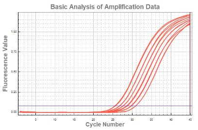
Excellent Reproducibility
Robust design ensures superior uniformity from run to run. Amplification curves for 96 replicates shown on a linear plot a logarithmic plot. Average quantification cycle (Cq)= 24±0.10.
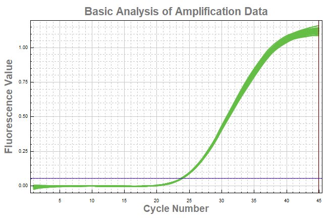
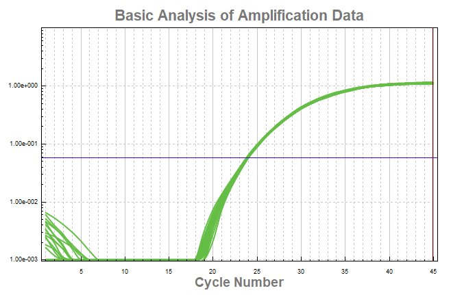
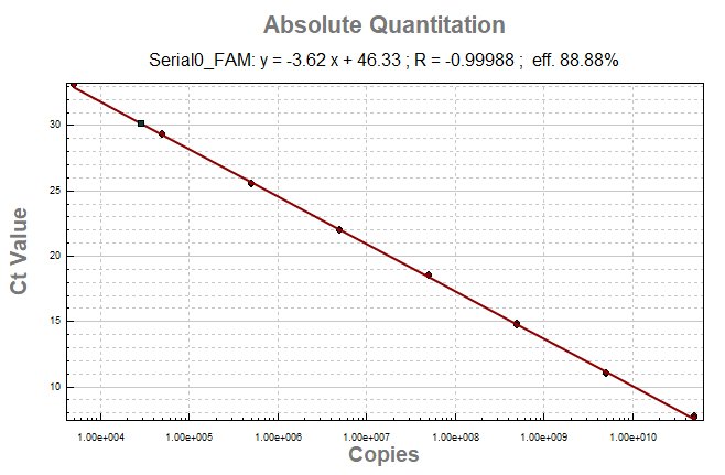
Up to 10-log dynamic range
Results on the nQ96-X4/5 System show excellent reproducibility and resolution down to very low copy numbers.
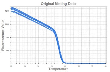
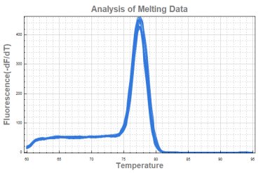
Melting Curve Analysis
96 replicates of human genomic DNA were amplified using SYBR reagent. The reactions were run under fast run mode, showing thermal uniformity as measured by the derivative peak with a melting temperature (Tm) of 77.5°C (standard deviation 0.05°C).
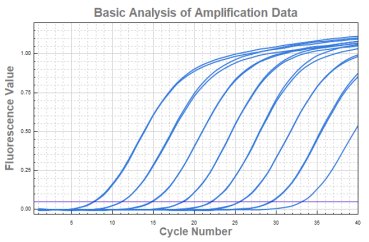
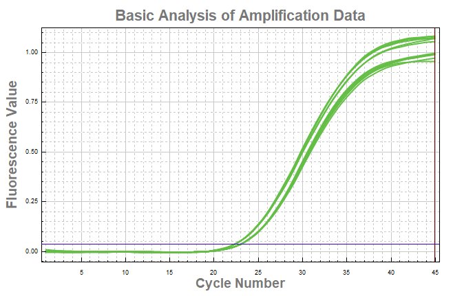
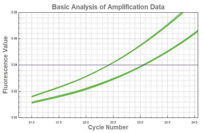
High Resolution
The excellent uniformity of temperature together with quick detection of data guarantee the reaction temperature and time to be identical between different tubes, so it can reliably detect 1.33-fold differences in target amount. The figure 1 shows the curve of 6 gradient dilutions: in 2X The figure 2 and 3 shows the curve of 1.5X dilutions.
specification
| Thermal Control System | |
| Sample capacity | 8*0.1mL PCR strip*12
96*0.1mL individual tube 0.1mL PCR Plate |
| Sample volume | 10-50 μL |
| Heating and Cooling method | Peltier |
| Maximum ramping rate | 6.0° C/s |
| Temperature range | 4-100 °C |
| Temperature accuracy | ± 0.2°C |
| Temperature uniformity | ±0.2°C@60°C ±0.3°C@95°C |
| Optical Detecting System | |
| Excitation light | 4/5pcs high-efficiency single color LED |
| Detector | SiPMT |
| Detection method | Time-resolved real-time scanning |
| Excitation/Emission wavelengths | 455-650nm/510-750nm |
| Detection Channels | 4 (optional 5 channels) |
| Supported dyes | FAM/SYBR Green, VIC/JOE/HEX/TET, ROX/Texas Red, Cy5/LIZ, (Optional- Cy5.5 for 5-channel model) |
| Multiplexing | Up to 4 targets (optional 5 targets for 5-channel model) |
| Sensitivity | 1 copy gene |
| Resolution | 1.33-fold copies difference in single-plex reaction |
| Dynamic range | 10 orders of magnitude |
| Analysis Mode | |
| Absolute quantification and melting curve analysis | |
| Data Export | |
| The original result, data and result in excel, program setting, amplification curve image | |